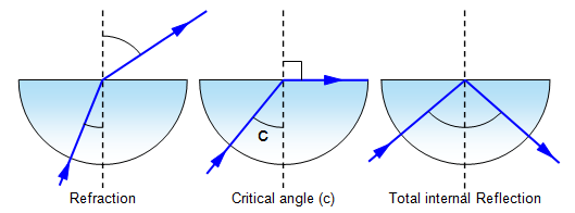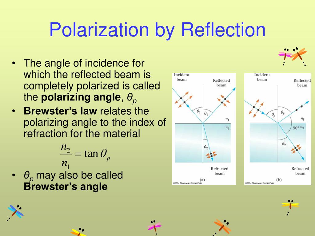

Scattering occurs when the width or lateral dimension of the tissue boundary is less than one wavelength (Animation 1.2.4). In animation 1.2.3, as the ultrasound wave passes through each tissue boundary, it loses some energy or amplitude while some of the wave is reflected. The difference in acoustic impedance between two tissues accounts for the amount of reflection that will occur at the tissue border. The change in impedance between two structures is call acoustic impedance mismatch. Large changes in density between two tissues will result in a large change of acoustic impedance. Acoustic impedance is determined by the density of the tissue. The amount of change of acoustic impedance will determine the amount of reflection. (Animation 1.2.2) Tissue boundaries that are smooth and have a width greater than the ultrasound wave act as a mirror or a specular reflector which results in a significant amount of reflection of the signal.Īs the ultrasound wave travels through one medium or tissue into another medium or tissue, a change in acoustic impedance occurs. If the tissue boundary width is less than the wavelength of the ultrasound wave, the ultrasound wave will not be reflected. In fact, the part of the parallel wall is not seen because the signal has been lost, which is commonly called dropout.īesides angle of incidence, the tissue boundary width impacts the amount of the reflected signal. Walls parallel to the ultrasound signal result in poor signal reflection. The walls parallel to the beam are not easily visualized. The myocardial walls that are perpendicular to the ultrasound beam act as excellent reflectors of the ultrasound signal. įor example, in Figure 1.2.1 and Video 1.2.1, the myocardial walls that are perpendicular to the ultrasound beam are easily visualized. When the ultrasound wave is not reflected, either due to a parallel tissue boundary or high acoustic impedance, the ultrasound signal is not recorded and the display shows a lack of signal or dropout of the ultrasound picture. Tissue boundaries that are perpendicular to the ultrasound wave's path act as excellent reflectors, whereas, tissue boundaries that are parallel to the ultrasound wave's path act as poor reflectors. Reflection occurs at tissue boundaries and tissue interfaces. For reflection, the angle of incidence is less than 90 degrees.

The angle of incidence is the angle of the ultrasound beam and the tissue plane.

The major factors affecting the amount of reflection are: Reflection occurs when the ultrasound wave is deflected towards the transducer. Ultrasound waves interact with tissue in four basic manners. Ultrasound waves, when they strike a medium, cause expansion and compression of the medium. Tissue interaction has also lead to the development of new technologies, such as automatic border detection. An understanding of the basic interactions of tissue with ultrasound provides the basis of avoiding errors and misdiagnosis. The interaction can cause measurement errors, artifacts, and poor picture quality. Even with the perfect echocardiographic machine, we are still left with the ultrasound interaction with tissues. The perfect echocardiographic machine would produce an infinitesimally small ultrasound beam, an incredible sweep speed and a uniform energy throughout its beam length.


 0 kommentar(er)
